
Multiparametrik Prostat MRI prostat bezi ve komşu dokuların görüntülenmesini sağlayan non-invaziv, maliyet etkin ve yaşam kalitesini olumsuz etkilemeyen bir ileri görüntüleme yöntemidir. Prostat kanseri yanısıra enfeksiyon, benign prostat hiperplazisi (BPH) veya doğumsal anomalilerin tanısında da önemlidir.
Prostat biyopsisi öncesinde kanser varlığını değerlendirmede, biyopsi sonucu negatif olsa dahi güçlü kuşku varsa, kanser tanısı almış ise büyüklüğü ve yayılımını değerlendirmede ve tedavi sonrası olası nükslerin ve tedavi etkinliğinin izlenmesinde kullanılır.
Prostat kanserlerinin bazıları zararsız,bazıları agresif seyirlidir. Görülme sıklığı yüksek olsa da; zararsız bazı prostat kanserlerinin tedavisi her zaman gerekli değildir ve tedavi edilmese dahi klinik olarak önemli problemlere neden olmaz.
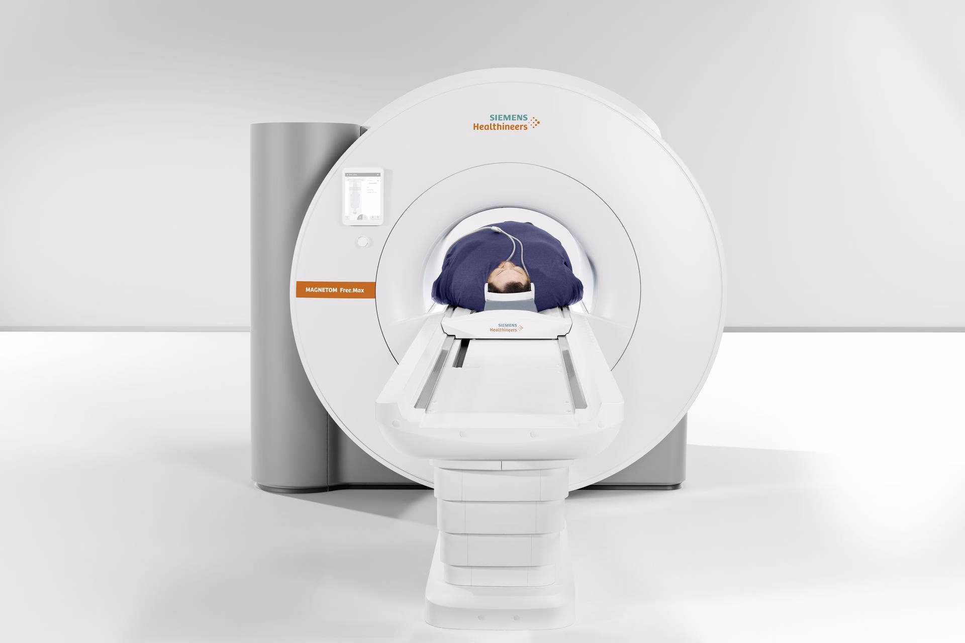
Multiparametrik MRI standart prostat MRI’a kıyasla çok daha ince detayları gösterir ve bu ileri teknoloji yöntemle prostat bezinin 3 boyutlu (3D) ve yüksek çözünürlüklü görüntüleri elde edilir.
Merkezlerimizde bulunan en gelişmiş ve ileri donanım ve yazılıma sahip 3 adet 3 Tesla MRI cihazı sayesinde çok hızlı randevu alabilirsiniz.
Neden İhtiyaç Duyulur?
- Biyopsi ihtiyacı olup olmadığının değerlendirilmesinde,
- Biyopsiye kılavuz olarak,
- Tedavi edilmeyen hastaların aktif izleminde,
- Kanserin büyüklüğü ve yayılımını saptamada,
- Doğumsal anomalilerde,
- Pelvis bölgesi ameliyatlarına bağlı komplikasyonların saptanmasında

Şimdi randevu almaya hazır mısınız?